Interventions

USG Guided Biopsy

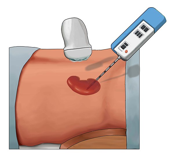
Our team of experienced radiologists and technicians provide USG (Ultrasound Guided) biopsies to help diagnose a wide range of conditions. Using real-time ultrasound imaging, we can precisely guide a biopsy needle to collect a small sample of tissue from the area of concern.

USG guided biopsies are commonly used to investigate:

  • Suspicious lumps or masses in the breast, thyroid, liver, kidney, or other organs
  • Fluid collections or abnormal growths
  • Bone and soft tissue lesions

The procedure is typically performed on an outpatient basis and only takes 30-60 minutes. We use local anesthesia to minimize discomfort, and patients can usually return to their normal activities the same day. Our state-of-the-art imaging equipment and specialized expertise ensure an accurate, safe, and comfortable biopsy experience.


CT Guided Biopsy

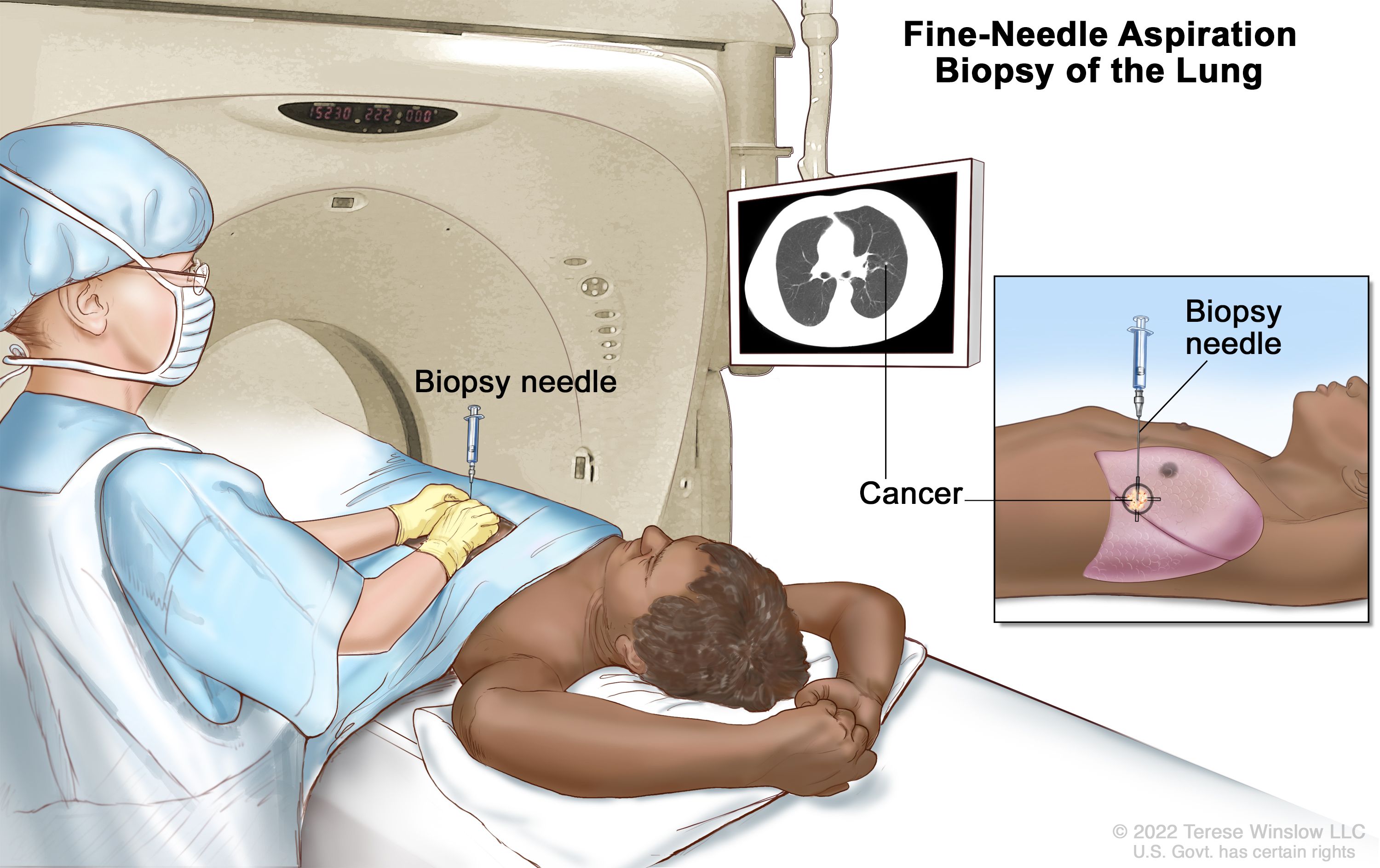
Our radiology team utilizes advanced CT (computed tomography) imaging to precisely guide biopsies and help diagnose a variety of medical conditions. Using real-time CT scans, we can accurately locate and sample tissue from hard-to-reach areas of the body.

CT guided biopsies are commonly used to investigate:

  • Lung nodules or masses
  • Liver, kidney, or adrenal lesions
  • Bone tumors and abnormalities
  • Lymph nodes and other deep-seated growths

The procedure is typically performed on an outpatient basis and takes around 30-60 minutes. We use local anesthesia to keep you comfortable, and most patients can return to their normal activities shortly after the biopsy. Our board-certified radiologists have extensive experience performing CT guided biopsies. We utilize the latest imaging technology and techniques to ensure an accurate diagnosis while minimizing any discomfort or complications.


FNAC (Fine Needle Aspiration Cytology)

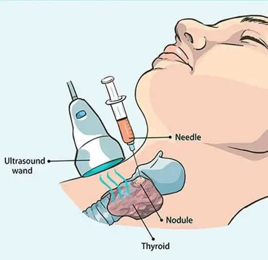
Fine needle aspiration cytology (FNAC) is a minimally invasive procedure used to collect cell samples from suspicious lumps, masses, or lesions. Our experienced radiologists and cytopathologists utilize specialized techniques and equipment to accurately obtain and analyze these cell samples.

FNAC is commonly used to investigate:

  • Thyroid nodules or growths
  • Breast lumps or suspicious areas
  • Lymph node enlargement
  • Lumps or masses in the neck, abdomen, or other areas

During the procedure, a thin, hollow needle is inserted into the area of concern under imaging guidance (such as ultrasound). The cells collected are then prepared and examined under a microscope to help determine the underlying cause. FNAC is a quick, safe, and relatively painless procedure that can often be performed on an outpatient basis. It provides valuable diagnostic information to guide further treatment planning.


Pigtail Insertion

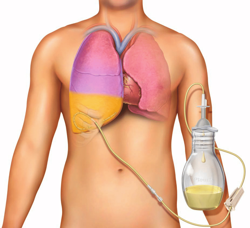
Our interventional radiology team specializes in the placement of pigtail catheters to drain fluid collections or relieve pressure buildup in the body. Using advanced imaging guidance, we can precisely insert these small, flexible tubes to effectively manage a variety of medical conditions.

Pigtail catheters are commonly used to:

  • Drain abdominal, pelvic, or thoracic fluid collections
  • Decompress obstructed urine flow from the kidneys
  • Drain infected or symptomatic cysts or abscesses
  • Manage malignant fluid buildup (such as pleural or ascitic effusions)

The procedure is typically performed on an outpatient basis using local anesthesia and mild sedation. Under imaging guidance, such as ultrasound or CT, the radiologist will make a small incision and insert the pigtail catheter into the target area. The procedure usually takes 30-60 minutes. Following the insertion, the catheter can remain in place for days or weeks to continuously drain fluid and relieve symptoms. Our team will provide detailed instructions on catheter care and monitoring.


Kidney Biopsy

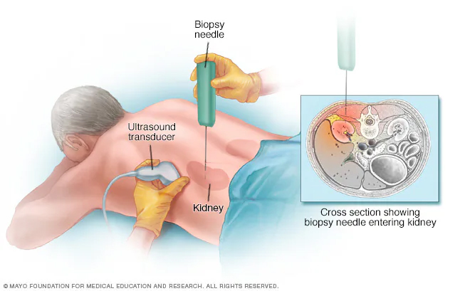
Our team of experienced radiologists performs kidney biopsies using advanced imaging guidance to collect tissue samples and help diagnose a variety of kidney conditions. This minimally invasive procedure allows us to obtain valuable information to guide your medical treatment.

Kidney biopsies are commonly used to investigate:

  • Unexplained changes in kidney function or laboratory test results
  • Suspected kidney disease, such as glomerulonephritis or nephrotic syndrome
  • Evaluation of kidney transplant function
  • Certain types of kidney tumors or masses

During the biopsy, a small needle is inserted through the skin and into the kidney under the guidance of real-time imaging, such as ultrasound or CT. This allows us to precisely target the area of interest and obtain a small sample of kidney tissue for analysis. The procedure is typically performed on an outpatient basis using local anesthesia. Most patients experience minimal discomfort and can return to their normal activities within a day or two.


Liver Abscess Drainage

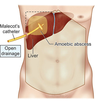
Our interventional radiology team specializes in the minimally invasive drainage of liver abscesses using advanced imaging guidance. This procedure helps evacuate infected fluid collections and promote healing, often avoiding the need for more invasive surgery.

Liver abscesses can develop due to bacterial, fungal, or parasitic infections, or as a complication of other medical conditions. Prompt drainage is often necessary to manage symptoms and prevent the spread of infection. Our radiologists utilize real-time imaging, such as ultrasound or CT scans, to precisely locate the abscess and guide a small drainage catheter into the affected area. This hollow tube is then left in place to continuously drain the infected fluid until the abscess has resolved.

The procedure is typically performed on an outpatient basis using local anesthesia and mild sedation. Most patients experience minimal discomfort and can return to their normal activities within a few days, with the catheter remaining in place for 1-2 weeks as needed. Our board-certified interventional radiologists have extensive experience in the safe and effective drainage of liver abscesses. We utilize the latest techniques and equipment to ensure optimal patient outcomes and minimize the risk of complications.


Permcath Procedure

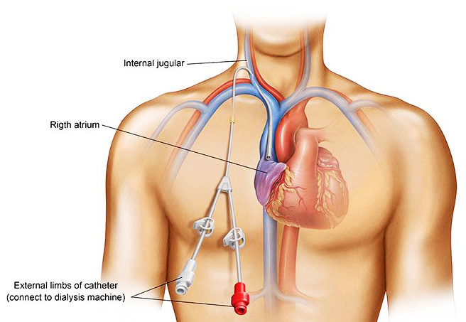
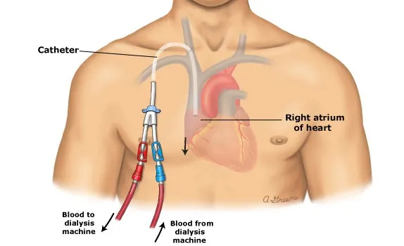
Our interventional radiology team specializes in the placement of permanent hemodialysis catheters, also known as Permcaths. This minimally invasive procedure provides reliable vascular access for patients requiring long-term dialysis treatment.

A Permcath is a small, flexible tube that is inserted into a large vein, typically in the neck or chest area. It allows easy and consistent access to the bloodstream for hemodialysis, eliminating the need for repeated needle sticks in the arm.

Our radiologists utilize advanced imaging guidance, such as ultrasound and fluoroscopy, to precisely insert the Permcath into the optimal vein location. The procedure is usually completed in under an hour using local anesthesia and mild sedation. Once the Permcath is in place, it can remain for months or even years, providing reliable vascular access for dialysis. Our team will provide detailed instructions on how to care for the catheter and monitor for any potential complications.

Central Line Procedure

Our expert interventional radiology team specializes in the placement of central venous catheters, also known as central lines. These specialized tubes provide reliable vascular access for a variety of medical treatments and procedures.

Central lines are commonly used to:

  • Administer medications, fluids, or nutrition directly into the bloodstream
  • Draw blood samples for testing
  • Provide access for hemodialysis or apheresis treatments
  • Monitor central venous pressure

Our radiologists utilize advanced imaging guidance, such as ultrasound and fluoroscopy, to carefully insert the central line into a large vein, typically in the neck, chest, or groin area. This minimally invasive procedure is usually completed in under an hour using local anesthesia. Once in place, the central line can remain for days, weeks, or even months, depending on the patient's needs. Our team will provide detailed instructions on how to properly care for the catheter and monitor for any potential complications.
Whatsapp icon